Every week, owners bring me hip radiographs and ask what I see. They have OFA certificates or PennHIP reports, but what they really want to know is whether the numbers and letters translate to a dog that will run without pain. After reading thousands of these films over twenty-five years, I have developed strong opinions about what matters and what gets overlooked.
The Positioning Problem
Before I say anything about hip quality, I look at positioning. A poorly positioned radiograph can make excellent hips look dysplastic or hide early pathology in what appears to be a normal joint. The OFA standard requires the dog in dorsal recumbency with femurs parallel and patellae centered over the trochlear grooves. Sounds straightforward until you try doing it with a nervous German Shepherd who would rather be anywhere else.
I see tilted pelves constantly. When the pelvis rotates even slightly, one acetabulum appears deeper than the other. The obturator foramina should be symmetrical. If they are not, the positioning is off, and the evaluation becomes unreliable. Understanding what hip dysplasia actually is helps owners interpret these findings more meaningfully. I have returned films to referral practices asking for repositioning. This makes me unpopular with some colleagues, but I would rather be accurate than polite.
Sedation quality matters enormously. Light sedation means muscle tension, and muscle tension can mask laxity. A dog with subluxating hips under light sedation may show reasonable coverage because the muscles are actively holding the femoral head in place. Deep sedation reveals the true relationship between bone and socket.
What OFA Grades Actually Measure
The Orthopedic Foundation for Animals uses a seven-point scale from Excellent to Severe. Three radiologists independently evaluate each submission and reach a consensus. This system has served breeding programs for decades, but it has limitations that owners should understand.

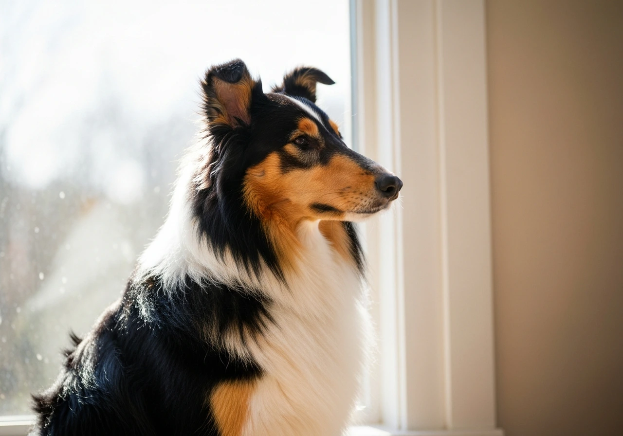
Excellent hips show deep acetabular coverage with the femoral head seated tightly. The Norberg angle exceeds 105 degrees, and there is no evidence of remodeling. These hips make me smile. In German Shepherds, I see perhaps one Excellent rating for every fifteen submissions. The breed's working structure, with that distinctive rear angulation, does not favor textbook hip conformation.
Good is where most healthy breeding dogs land. The femoral head sits well within the acetabulum with minor incongruence. I have operated on zero dogs with Good hips for dysplasia-related pain. Zero. If your breeding dog has Good hips, that is genuinely good news.
Fair is where the trouble starts in terms of interpretation. Fair hips show more incongruence but remain within normal limits. Some Fair hips will never cause problems. Others will develop arthritis by age five. The OFA grade alone cannot predict which path your dog will take.
The Grades I Worry About
Borderline designation frustrates everyone. The radiologists could not reach consensus, so they punt. I understand why this category exists, but it leaves owners in limbo. My advice for Borderline results is always the same: wait six months and resubmit. Skeletal maturity continues until roughly two years in large breeds, and a Borderline at eighteen months sometimes clarifies to Fair or Mild by twenty-four months.
Mild dysplasia describes hips with shallow acetabula or early subluxation. The femoral head is not seated deeply, but there is no significant arthritis yet. Many Mild dogs live comfortable lives, particularly if we manage their weight aggressively. I do not recommend breeding Mild dogs, but I have seen Mild hips function well into double digits with proper management.
Moderate changes my conversation entirely. Moderate means obvious subluxation, often with secondary arthritic changes beginning. These dogs may already have discomfort that owners attribute to normal slowing down. When I see Moderate dysplasia in a young dog, we need to discuss surgical intervention seriously.
Severe dysplasia is unmistakable even to untrained eyes. Complete luxation, flattened femoral heads, severe osteoarthritis. These hips have failed catastrophically. I operate on Severe hips weekly, and the transformation after surgery can be remarkable, but the joint itself is beyond salvage. We are replacing, not repairing.
The Ortolani Test: What X-Rays Cannot Show
Static radiographs capture one moment in time with the dog positioned artificially. They cannot assess dynamic laxity, which is often the first sign of developing dysplasia. The Ortolani test fills this gap, and I perform it on every Shepherd puppy I examine.
With the dog sedated and relaxed, I apply medial pressure to the stifle while simultaneously pushing the femur dorsally. In a normal hip, nothing happens. The femoral head stays put. In a dysplastic hip, the femoral head subluxates out of the acetabulum. Then, as I abduct the limb, you feel a distinct clunk as the head reduces back into position. That clunk is the Ortolani sign, and it tells me more about future problems than the radiograph on my viewer.
Puppies as young as sixteen weeks can show positive Ortolani. At that age, the radiograph may look entirely normal because the secondary changes have not developed. If I feel that clunk in a young Shepherd, we need to discuss PennHIP evaluation and potentially juvenile pubic symphysiodesis or triple pelvic osteotomy before the window closes.
PennHIP Versus OFA: Different Questions, Different Answers
PennHIP measures distraction index, essentially how far the femoral head can be pulled from the acetabulum under standardized force. This quantifies laxity on a continuous scale rather than categorical grades. A distraction index of 0.3 puts a dog in the tightest-hipped 30% of its breed. An index of 0.7 means significant laxity and high dysplasia risk.
I recommend PennHIP for breeding dogs and for puppies where early intervention might be considered. The continuous scale allows more nuanced comparisons, and the breed-specific percentile rankings help contextualize results. A German Shepherd with a DI of 0.45 sits around the median for the breed, neither particularly tight nor loose. The same DI in a Border Collie would be concerning, as that breed typically shows tighter hips.
For pet owners primarily concerned with whether their dog will need surgery, OFA evaluation at twenty-four months often suffices. The categorical grades are easier to interpret, and by that age, the clinical picture usually matches the radiographic findings.
Secondary Changes I Look For
Beyond acetabular depth and femoral head coverage, several radiographic signs concern me:
- Morgan line: A curvilinear opacity along the caudal femoral neck. This represents early enthesophyte formation and predicts progressive osteoarthritis. I see Morgan lines in dogs with otherwise Fair hips, and it changes my prognosis.
- Femoral head remodeling: The normal spherical head becomes flattened or mushroomed as the cartilage breaks down. Once remodeling starts, it does not reverse.
- Acetabular filling: The body tries to compensate for a shallow socket by building new bone. This osteophyte formation initially improves coverage but eventually creates mechanical impingement and pain.
- Subchondral sclerosis: Increased bone density beneath the cartilage surface indicates chronic stress and early arthritis even when joint space appears preserved.
The Frustration of Late Diagnosis
I receive referrals constantly for dogs who have been limping for months before anyone took radiographs. The owners assumed the dog was tired or getting older. By the time I see them, we are looking at advanced arthritic changes and limited options.
Early screening saves dogs from preventable suffering. A Shepherd puppy with lax hips identified at four months has surgical options that disappear by eighteen months. A young adult with Mild changes benefits from aggressive weight management and physical therapy before arthritis establishes. Waiting until the dog cannot rise comfortably means we have already lost critical intervention windows.
If you own a German Shepherd, Belgian Malinois, or any herding breed with known dysplasia prevalence, get baseline radiographs. Even if the dog moves beautifully. Even if both parents had OFA clearances. The genetics of hip dysplasia are polygenic, and two cleared parents can absolutely produce an affected puppy. Knowing early lets us plan intelligently.
What I Tell Owners After Reviewing Films
My approach depends on what I see and what the owner needs to hear. For Excellent or Good hips, I congratulate them and recommend repeat evaluation only if clinical signs develop. For Fair hips, I discuss weight monitoring and suggest annual recheck radiographs to track any progression.
For dysplastic hips, the conversation becomes more detailed. I explain what the grade means functionally, outline the likely progression, and present the realistic options. Some owners want immediate surgical consultation. Others prefer to start with conservative management. Both approaches can be appropriate depending on the dog's age, activity level, and current comfort. For breeders, I also recommend evaluating elbows alongside hips, since joint scoring should be comprehensive. Resources like the elbow score database can help track elbow evaluations across breeding lines.
What I never do is minimize the finding. Telling an owner their dog has moderate dysplasia but will probably be fine is unfair to both owner and dog. That hip will cause problems. The only questions are when and how severe. I would rather have a difficult conversation now than perform emergency surgery on a dog in crisis later.
For breeders seeking to integrate OFA results into a broader selection strategy, our guide on breeding selection and genetic testing explains how to combine radiographic findings with estimated breeding values and genomic data. For comprehensive information on canine orthopedic genetics and testing, the genetic testing resources at The Herding Gene provide additional context for breeders evaluating their programs.