Medical Imaging: What It Is and How It Works
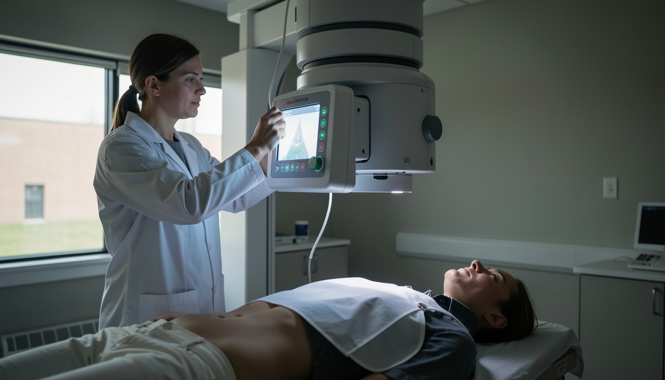
Medical imaging is the use of X-rays, CT, MRI, ultrasound, and other techniques to create pictures of the inside of the body. Each examination is ordered to answer a specific clinical question, and the type of image is chosen based on what needs to be seen. Understanding how medical imaging works helps patients and owners follow the process from referral to result.
What is medical imaging?
Medical imaging covers several methods that produce pictures of internal structures. X-rays use ionising radiation to show bones and dense tissue. CT scans combine multiple X-ray images to create cross-sectional views. MRI uses magnetic fields and radio waves to show soft tissues. Ultrasound uses high-frequency sound waves to show organs and fluid. Each method has strengths and limits, and the choice depends on the clinical question.
A clinician asks a question first, such as whether a bone is fractured or whether a joint is inflamed. The imaging examination is a picture taken to answer that question. The referrer, usually a doctor or vet, decides that an image is needed. The practitioner, often a radiographer or radiologist, carries out the examination. The operator uses the equipment, and the employer ensures the rules are followed. These four duty holders are defined in regulation 2 of the Ionising Radiation (Medical Exposure) Regulations 2017. For a plain explanation of how medical imaging works, uk-radiology.co.uk describes each modality and the rules that govern its use.
How is a medical image made?
Making an image starts with a question from a clinician. The referrer justifies the exposure, meaning they confirm that the benefit outweighs the risk. For X-rays and CT, the operator positions the patient and sets the machine. The radiation passes through the body and is captured by a detector. For MRI, the patient lies inside a strong magnetic field while radio waves disturb hydrogen atoms, and the signals are turned into images. For ultrasound, a probe sends sound waves and receives echoes. The resulting pictures are reviewed by a practitioner who reports the findings.
Who decides that an image should be made?
The decision follows a legal framework. The referrer, a clinician, asks the clinical question and refers the patient. The practitioner, a trained professional, justifies the exposure and authorises it. The operator, who may be a radiographer, physically performs the examination. The employer provides the equipment, training, and protocols. This separation of duties ensures that each exposure has a clear clinical purpose. The Ionising Radiation (Medical Exposure) Regulations 2017 set out these roles in UK law.
What are the main imaging modalities?
X-ray is the oldest and most common. It uses a small dose of radiation and is fast. CT provides detailed cross-sections and is often used in emergencies. MRI gives excellent soft-tissue contrast without ionising radiation. Ultrasound is real-time, portable, and used for abdominal, cardiac, and musculoskeletal scans. Each modality has a different physical basis, and the referrer selects the one that best answers the question.
How much radiation is involved?
Doses vary by examination. A chest X-ray delivers about 0.014 mSv. The average natural background dose in the UK is 2.7 mSv per year. These figures come from the UK Health Security Agency comparison table, published on 18 March 2011. CT scans involve higher doses than plain X-rays, while MRI and ultrasound use no ionising radiation. The referrer weighs the benefit of the image against the radiation risk.
What is the history of medical imaging?
X-rays were discovered in 1895. The Roentgen Society, the first X-ray association, was founded in 1897, as published by the Royal College of Radiologists. Since then, CT, MRI, and ultrasound have been developed. Each new method expanded what clinicians can see without surgery. The basic principle remains: an image answers a question that a clinician has already asked.
How does imaging help in practice?
In human medicine, imaging confirms fractures, detects tumours, and guides procedures. In veterinary medicine, it assesses hip dysplasia, fractures, and soft-tissue injuries. For shepherd breeds, hip X-rays are common to screen for dysplasia. The same rules apply: a vet refers, a radiographer or vet takes the image, and a radiologist reports. The image guides treatment decisions.
What should a patient or owner expect?
A patient or owner can expect a referral from a clinician, an appointment for the scan, and a report sent back to the referrer. The examination is usually painless, though some require stillness or contrast agents. The results are explained by the referrer. Understanding the process reduces anxiety and helps people ask informed questions about their care.